SWITCH2017: Stroke Workshop on Imaging and Treatment CHallenges

Recent Updates

Overview

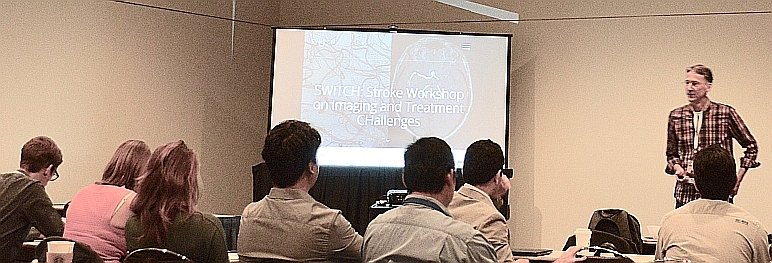
SWITCH is a half-day workshop focused on imaging related to stroke diagnosis and treatment. The main goals of the workshop are 1) to introduce the clinical background of challenges/opportunities related to imaging for stroke that are relevant for researchers working in the MICCAI field, and 2) to stimulate discussion and ideas exchange. To this end, there will be keynotes by clinical experts in stroke imaging and treatment, as well as presentations by researchers of on-going work. The tutorial character of the keynotes makes this workshop a perfect introduction for the ISLES workshop in the afternoon.

Call for papers

SWITCH is soliciting manuscripts that contribute to the workshop, addressing among others one of the following subjects:

  • Novel image processing approaches for lesion detection and quantification
  • Imaging based approaches to treatment decision making
  • Image guidance during interventions
  • Imaging based quantification of treatment

We encourage work involving, among others, the following imaging modalities:

  • CT-variants (CT, CTA, CTP, multi-phase CTA)
  • MR (DWI)
  • X-ray

Submissions should follow the LNCS guidelines for formatting, and consists of full papers of at most 8 pages.

All submitted manuscripts will be reviewed by the organizing committee on applicability to the workshop topic, scientific quality and clinical relevance. Note that we value novel methodology as well as thorough evaluation. Accepted manuscripts will get an oral presentation during the workshop at MICCAI. The SWITCH half-day workshop will join the MICCAI initiative for bundled joint Sep 2017: LNCS Proceedings of the satellite events.

Related Workshops

Important Dates

  • March 30, 2017 Website up
  • June 19, 2017 Paper Submission Deadline (Extended from June 5)
  • July 12, 2017 Paper Decision
  • July 24, 2017 Camera Ready Deadline
  • July 24, 2017 Full Program Available
  • September 10, 2017 Workshop

Tentative Program

  • 08:00 - 08:10 Introduction
  • 08:10 - 09:00 Keynote: Roland Wiest, Support Center of Advanced Neuroimaging, Inselspital Bern
    Neuroimaging of stroke: A role of MRI · abstract
  • 09:00 - 09:50 Keynote: Kambiz Nael, Mount Sinai Hospital, New York
    CT imaging in care of acute stroke patients · abstract
  • 10:00 - 10:30 Refreshments
  • 10:30 - 11:20 Keynote: Vitor Mendes Pereira, University of Toronto
    Mechanical thrombectomy and periprocedural imaging techniques · abstract · bio
  • 11:20 - 12:00 Presentations by authors of accepted papers

Details: About SWITCH

Stroke

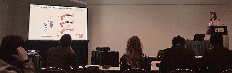
Stroke, as a result of insufficient blood supply to the brain, is the 4th cause of death and disability world-wide. The causes of stroke are either a bleeding (hemorrhage) in the brain, or an occlusion of a vessel feeding (part of) the brain. In the latter case, the occlusion may originate from a brain vessel itself, or be a thrombus that originates from a more proximal location (heart, carotid artery).

Imaging

Currently patients generally undergo non-contrast CT imaging, as well as a contrast-enhanced protocol: single phase CTA, multiphase CTA, or CT Perfusion. Some studies also involve MR imaging. From these images, information such as lesion and lesion size, composition of the thrombus, collateral flow and perfusion of brain tissue may be obtained, but few validated tools and quantitative imaging biomarkers have been developed and evaluated for clinical use so far. Often, subjective and at best semi-quantitative measures (e.g. ASPECT score for ischemic stroke) based on the CT images are being used to select patients and to decide on treatment strategy. A recent study that discussed 5 large clinical trials regarding stroke treatment concluded that “it is time to evaluate the optimal imaging protocol for hyper-acute stroke”.

Goals

Therefore, the main purposes of this Workshop are

  1. To introduce the challenges/opportunities related to imaging for stroke (assessment and diagnosis, therapy decision making, therapy guidance, therapy assessment, outcome prediction) that are relevant for researchers working in the “MICCAI” field. This will be done via clinical keynote speakers.
  2. To get informed on current work in this field. This will be done via presentations of the submitted contributions.
  3. To bring together researchers in this field, and stimulate further ideas exchange: via the discussions and interaction during the whole workshop.

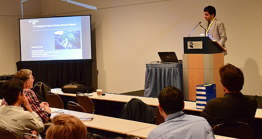
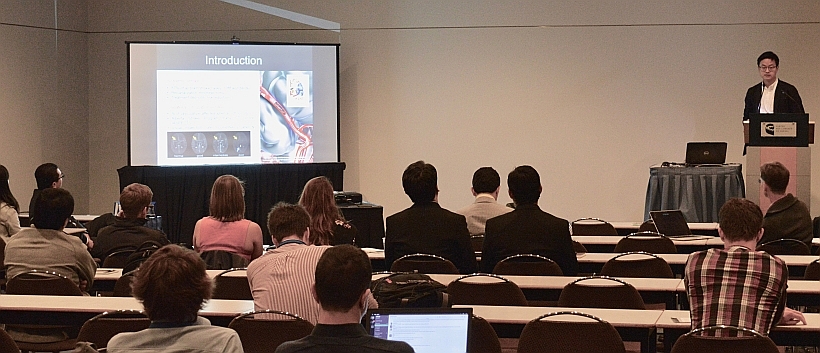
Thus, we intend to compose a program with presentations by researchers of on-going work, based on submitted manuscripts as well as key-note presentations by clinical experts in stroke management, modern imaging for stroke.

Challenges

Challenges in stroke imaging that will be addressed by the keynotes are:

  1. What information is required to determine the proper treatment for ischemic stroke patients? What underlying mechanisms are relevant (properties of the thrombus, collaterals, CBF, CBV, …), what (imaging) methods are best to get the appropriate information? Is CT(A) sufficient, what about CT perfusion, is MR required, can we also use rotational X-ray (potentially yielding a time gain, imaging the patient in the intervention room)? Next to these more fundamental questions, there are also more pragmatic (logistics) issues: CT is generally available, MR or an angio room availability is less common.
  2. How can we contribute to large studies investigating the above questions? What quantifications/biomarkers should be computed, from what modality?
  3. How can we optimally support the treatment of the patient (image guidance in thrombectomy)? The thrombus (and distal vessels) are hard to visualize during the intervention. Are there ways to provide better guidance? Are there good ways to assess whether the treatment is successful (and sufficient)? Can we predict patient outcome after the treatment?

Photos


SWITCH Organizing Committee

Theo van Walsum

Biomedical Imaging Group Rotterdam, Erasmus MC

Roman Peter

Biomedical Imaging Group Rotterdam, Erasmus MC

Wiro Niessen

Delft University of Technology, Delft and
Biomedical Imaging Group Rotterdam, Erasmus MC

Adrian Dalca

CSAIL, MIT and
MGH, Harvard Medical School

Mauricio Reyes

University of Bern
Biomedical Neuroimage Analysis Group

Roland Wiest

Support Center of Advanced Neuroimaging, Inselspital Bern

Bart Emmer

Department of Radiology, Amsterdam Medical Center

Contact

Please contact us for further questions and comments via email at switch@miccai2017.org

Sponsors